Invisalign® as low as $3,379!

23
Jun
Castlebury Dental
Oral surgery can feel intimidating, especially if you’re unsure what to expect. Whether you need a wisdom tooth extraction, dental implant placement, or another surgical procedure, it’s natural to have questions about safety and recovery. Fortunately, modern dental technology has transformed the way oral surgery is planned and performed. One of the most valuable advancements is digital imaging, which gives dentists a detailed view of your teeth, jaw, nerves, and surrounding structures before treatment even begins.
Instead of relying solely on traditional X-rays, digital imaging provides highly detailed images that help dentists diagnose conditions accurately, create personalized treatment plans, and reduce the risk of unexpected complications. At Castlebury Dental in Eagle, ID, advanced digital imaging technology plays an important role in delivering precise, comfortable, and predictable oral surgery for patients of all ages.
Let’s explore how this technology improves both safety and outcomes during oral surgery.
What Is Digital Imaging in Dentistry?
Digital imaging refers to modern imaging technologies that capture detailed pictures of your teeth, jawbone, soft tissues, and surrounding oral structures. Unlike traditional film X-rays, digital images are available almost instantly, allowing your dentist to evaluate your oral health in real time.
Depending on your treatment needs, digital imaging may include:
- Digital dental X-rays
- Panoramic imaging
- Three-dimensional cone beam computed tomography (CBCT)
- Intraoral digital photographs
Each type of imaging provides valuable information that helps your dentist understand your unique anatomy before recommending surgery.
Because the images are clear and highly detailed, they support more accurate diagnoses and better-informed treatment decisions.
Why Careful Planning Matters Before Oral Surgery
Successful oral surgery begins long before the actual procedure. Every patient’s mouth is different, and important structures such as nerves, blood vessels, sinuses, and bone density vary from person to person.
Without detailed imaging, these anatomical differences may not be fully visible.
Digital imaging allows your dentist to carefully evaluate:
- Tooth position
- Bone quality and volume
- Jaw structure
- Root shape
- Nearby nerves
- Sinus location
- Existing infections
- Hidden fractures or abnormalities
By identifying these factors beforehand, your dental team can plan the safest approach while minimizing unnecessary risks.
How Digital Imaging Improves Surgical Accuracy
Precision is one of the greatest advantages of digital imaging.
High-resolution images allow your dentist to measure distances, evaluate angles, and determine the ideal surgical approach before treatment begins.
For example, when placing a dental implant, your dentist can identify exactly where sufficient bone exists while avoiding important anatomical structures. During wisdom tooth removal, imaging helps determine whether tooth roots are close to major nerves or positioned in unusual directions.
This level of planning allows surgery to proceed more smoothly while reducing surprises during the procedure.
Protecting Important Nerves and Surrounding Structures
One of the primary goals during oral surgery is protecting nearby nerves, blood vessels, and healthy tissues.
Digital imaging helps dentists identify the precise location of structures that cannot always be seen during a clinical examination alone.
This is especially valuable when treating:
- Impacted wisdom teeth
- Dental implant cases
- Bone grafting procedures
- Surgical tooth extractions
Knowing exactly where these sensitive structures are located helps reduce the risk of accidental injury, numbness, excessive bleeding, or other complications.
Earlier Detection of Hidden Dental Problems
Not every dental issue is visible during a routine examination.
Digital imaging often reveals problems hidden beneath the surface, including:
- Bone infections
- Impacted teeth
- Tooth fractures
- Bone loss
- Cysts
- Abscesses
- Root abnormalities
Identifying these conditions before surgery allows your dentist to adjust the treatment plan if necessary and address additional concerns before they become larger problems.
Early diagnosis often leads to more successful outcomes and smoother healing.
Better Treatment Planning for Dental Implants
Dental implant placement requires careful precision because the implant must integrate securely with the jawbone.
Digital imaging allows your dentist to evaluate:
- Bone thickness
- Bone height
- Bone density
- Implant positioning
- Bite alignment
With three-dimensional imaging, your dentist can virtually plan implant placement before surgery even begins.
This advanced planning helps improve dental implant stability, supports long-term success, and reduces the likelihood of complications.
Making Wisdom Tooth Removal Safer
Wisdom teeth often develop in unpredictable ways. Some remain trapped beneath the gums, while others grow sideways or press against neighboring teeth.
Digital imaging helps determine:
- Whether wisdom teeth are impacted
- Root development
- Tooth orientation
- Proximity to the lower jaw nerve
- Potential effects on adjacent teeth
This information allows your dentist to choose the safest removal technique while reducing the risk of nerve injury and unnecessary trauma to surrounding tissues.
Lower Radiation with Modern Digital Technology
Many patients worry about radiation exposure during dental imaging.
Fortunately, digital imaging typically exposes patients to significantly less radiation than traditional film-based X-rays while producing clearer images.
Modern equipment captures high-quality images quickly, reducing the need for repeated exposures.
Your dentist only recommends imaging when it provides important information that benefits your diagnosis and treatment planning.
Helping Patients Understand Their Treatment
Digital imaging doesn’t only benefit the dental team—it also helps patients better understand their own oral health.
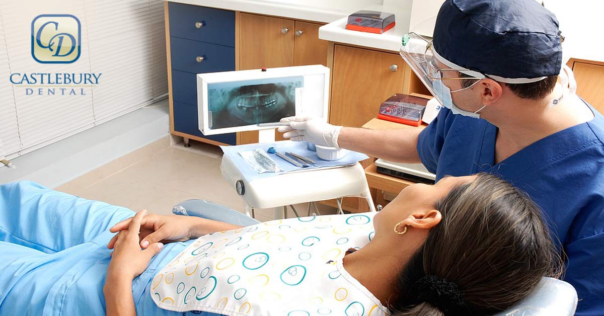
Instead of simply describing a problem, your dentist can display detailed images on a monitor and explain exactly what is happening.
Patients can often see:
- Impacted teeth
- Bone loss
- Infections
- Damaged roots
- Areas requiring treatment
Seeing these images often makes treatment recommendations easier to understand and allows patients to feel more confident about moving forward with oral surgery.
Clear communication helps reduce anxiety and encourages informed decision-making.
Supporting Faster Recovery
A well-planned surgery often leads to a smoother recovery.
Because digital imaging allows your dentist in Eagle, ID to work more precisely, procedures may involve less disruption to healthy tissues.
Benefits may include:
- Reduced swelling
- Less discomfort
- Shorter surgical time
- Improved healing
- Lower risk of complications
- More predictable results
Every patient’s healing process is unique, but careful planning plays an important role in supporting successful recovery.
Is Digital Imaging Safe for Most Patients?
Digital imaging is considered safe for most individuals, including children when clinically appropriate. Your dentist follows established safety guidelines and recommends imaging only when it provides valuable diagnostic information.
Protective measures are used whenever necessary, and the amount of radiation is kept as low as reasonably achievable while still obtaining high-quality images.
If you are pregnant or believe you may be pregnant, let your dentist know before imaging so appropriate precautions can be discussed.
Why Experience and Technology Work Together
Advanced technology is only one part of successful oral surgery. The experience of your dental team is equally important.
Digital imaging provides detailed information, but your dentist uses clinical expertise to interpret those images, develop an individualized treatment plan, and perform surgery with precision.
Combining modern technology with skilled care helps deliver safer procedures, improved comfort, and better long-term outcomes.
Experience Advanced Dental Technology at Castlebury Dental
Digital imaging has transformed modern oral surgery by making diagnosis more accurate, treatment planning more precise, and procedures safer for patients. From identifying hidden concerns to protecting important anatomical structures, this technology helps reduce risks while improving overall treatment outcomes.
At Castlebury Dental, located at 3209 W. Bavaria St., Eagle, ID 83616, we use advanced digital imaging technology to provide personalized care and carefully planned oral surgery. Whether you need a tooth extraction, dental implant consultation, or another surgical procedure, our team is committed to making your experience as safe and comfortable as possible.
Schedule an appointment today to learn how modern dental technology can help protect your oral health and support a successful treatment experience.
Frequently Asked Questions
Digital imaging uses advanced technology to create detailed images of your teeth, jaw, and surrounding structures, helping dentists diagnose problems and plan treatment with greater accuracy.
Yes. Digital imaging helps your dentist identify nerves, bone structure, impacted teeth, and other important anatomical features before surgery, allowing for more precise treatment planning.
Digital imaging provides clearer images almost instantly and typically uses less radiation than traditional film X-rays, making it a valuable tool for diagnosis and treatment planning.
It helps evaluate bone density, bone volume, and ideal implant placement, increasing the accuracy of the procedure and supporting long-term implant success.
Not always. Your dentist will recommend the type of imaging that best suits your specific condition and treatment needs to ensure safe and effective care.